When Skin Disease Isn’t Just Skin Disease: Recognising Epitheliotropic Lymphoma in Dogs and Cats
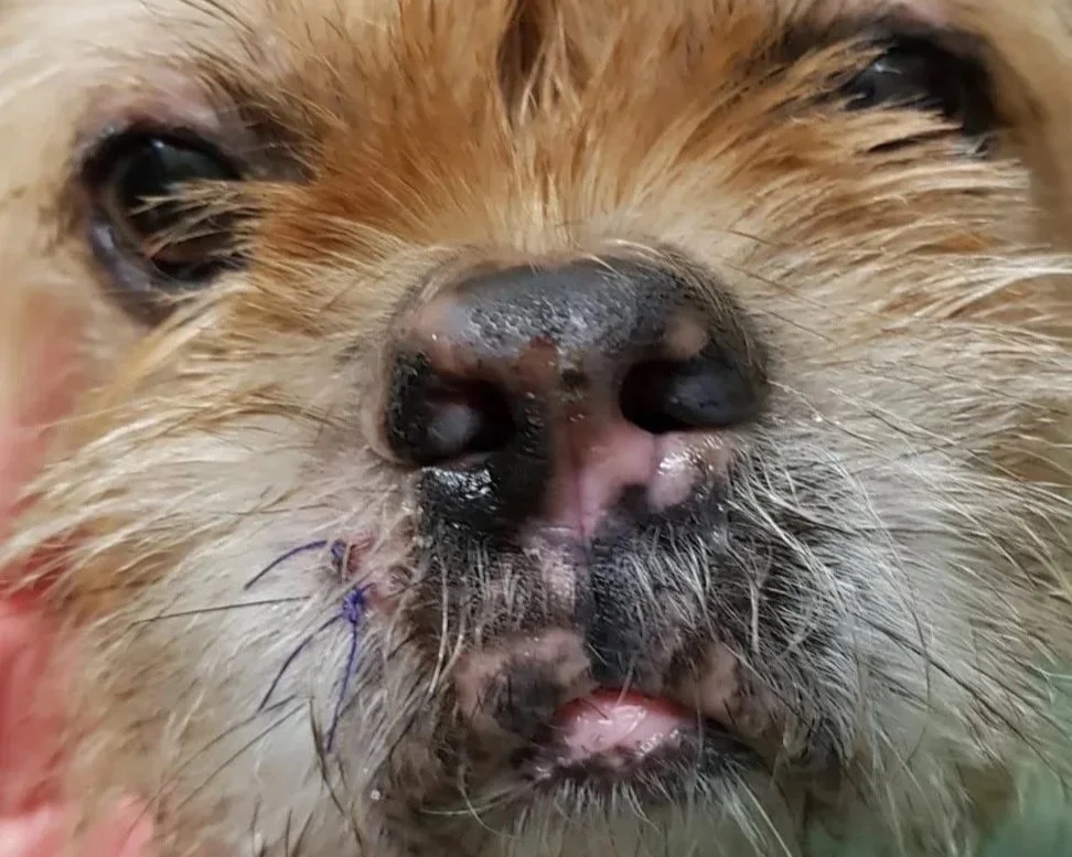
Epitheliotropic lymphoma is one of those cancers that can be easy to miss in its early stages.
Rather than presenting as an obvious tumour, it can initially look like an ordinary skin problem: redness, scaling, hair loss, itching, crusting or recurrent infections.
For owners, this can mean months of treating what appears to be dermatitis. For veterinarians, it can mean navigating a long list of dermatological differentials before the underlying diagnosis becomes apparent.
Epitheliotropic lymphoma is a form of cutaneous lymphoma in which neoplastic lymphocytes have a particular tendency to migrate into the epidermis and, in many cases, the hair follicle and adnexal structures. It is most commonly a T-cell lymphoma and is also referred to as cutaneous epitheliotropic T-cell lymphoma (eCTCL). Histologically, it encompasses patterns including mycosis fungoides and pagetoid reticulosis, while more extensive disease involving circulating neoplastic cells is associated with Sézary syndrome.
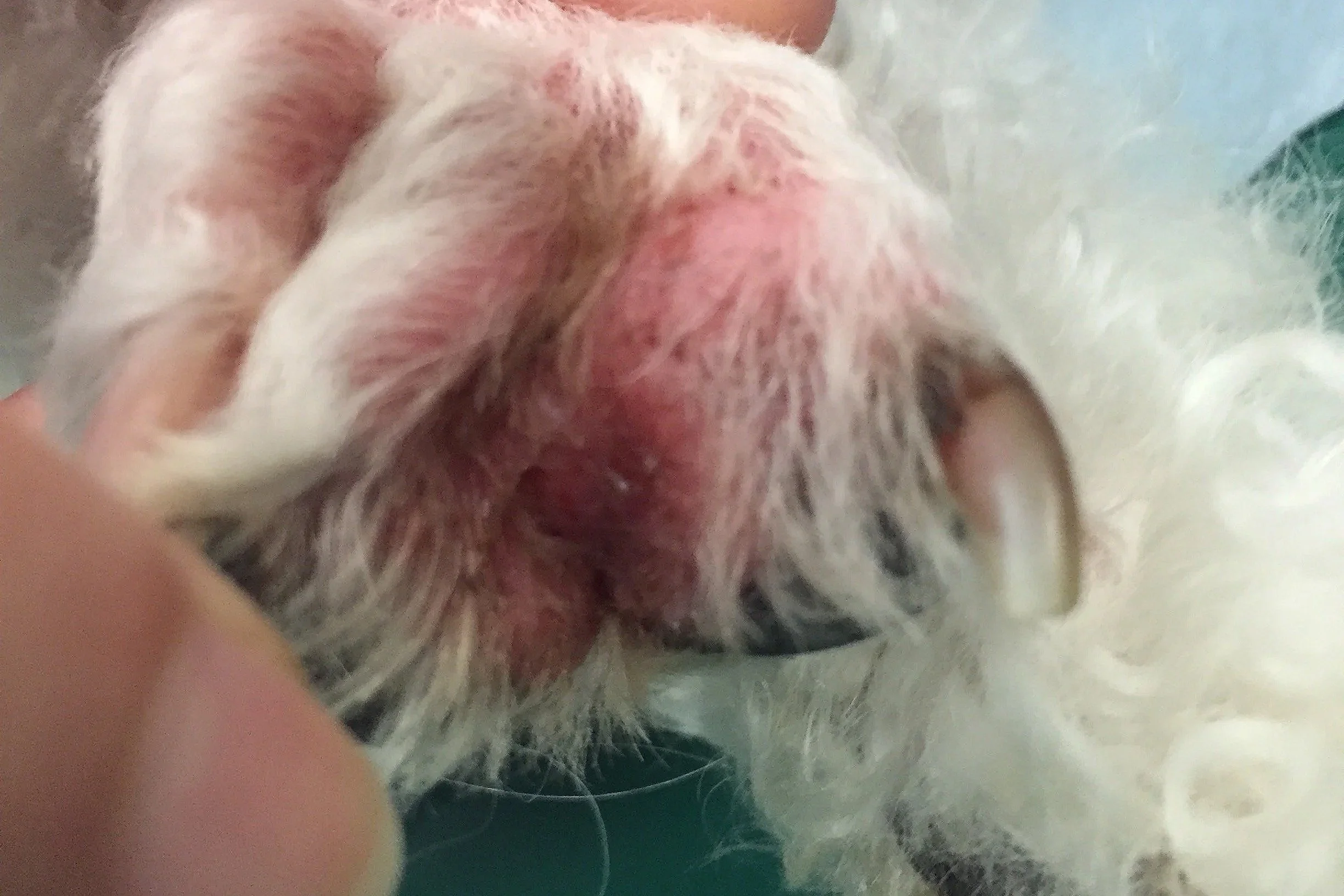
The dog with “chronic dermatitis”
A middle-aged to older dog presents with a history of gradually worsening skin disease.
Initially, the owner noticed:
Red, inflamed skin
Scaling
Hair thinning
Increasing itchiness
Recurrent areas of bacterial or yeast infection
The dog had been treated at various times with antibiotics, antifungals, anti-inflammatory medication and treatment for presumed allergic skin disease.
There were periods of improvement, but the disease always returned.
On closer examination, the distribution and character of the lesions were unusual. There were multifocal areas of erythema, alopecia and scaling, with some lesions becoming thicker and more plaque-like.
Skin biopsy revealed an infiltrate of atypical lymphocytes with epitheliotropism, involving the epidermis and follicular epithelium. Immunohistochemistry supported a T-cell phenotype, and clonality testing was consistent with a clonal T-cell population.
The diagnosis was cutaneous epitheliotropic T-cell lymphoma.
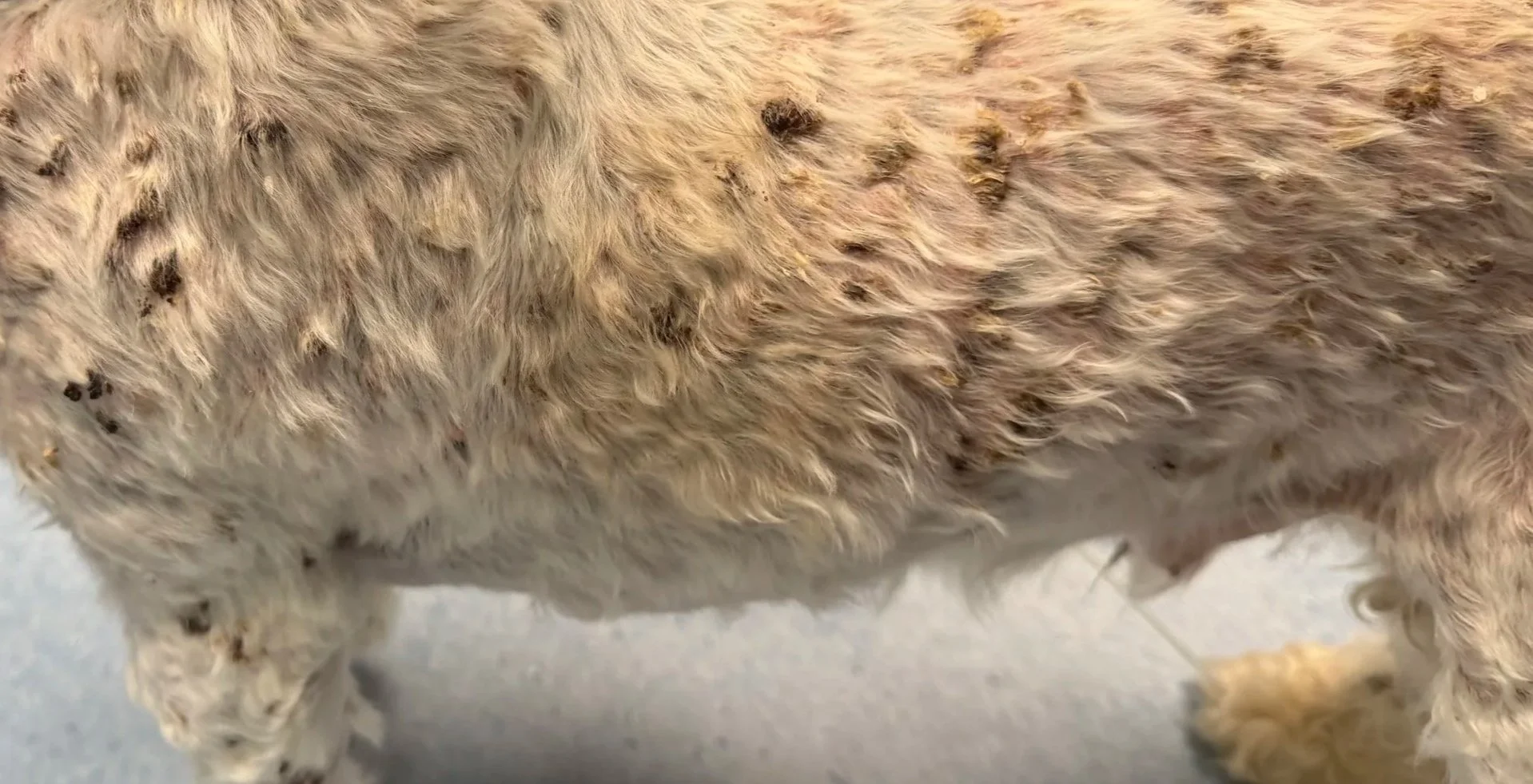
The dog with patches, plaques and pigment changes
Another presentation can be much more subtle.
The owner notices several areas of:
Hair loss
Redness
Scaling
Hyperpigmentation or hypopigmentation
Thickened skin
The lesions may initially resemble chronic inflammatory skin disease.
There may be no obvious mass.
In fact, in some dogs the disease looks more like a multifocal or generalised inflammatory skin disorder than cancer.
Published cases demonstrate just how variable the clinical appearance can be. In one series of 30 dogs with cutaneous epitheliotropic T-cell lymphoma, diffuse erythema was reported in 86.6% of cases, scaling in 60% and focal hypopigmentation in 50%. Mucocutaneous involvement occurred in approximately half of the dogs.
This is why the distribution and progression of the disease matter.
A lesion that is:
persistent → progressive → unusual → poorly responsive to conventional therapy
should prompt reconsideration of the diagnosis.
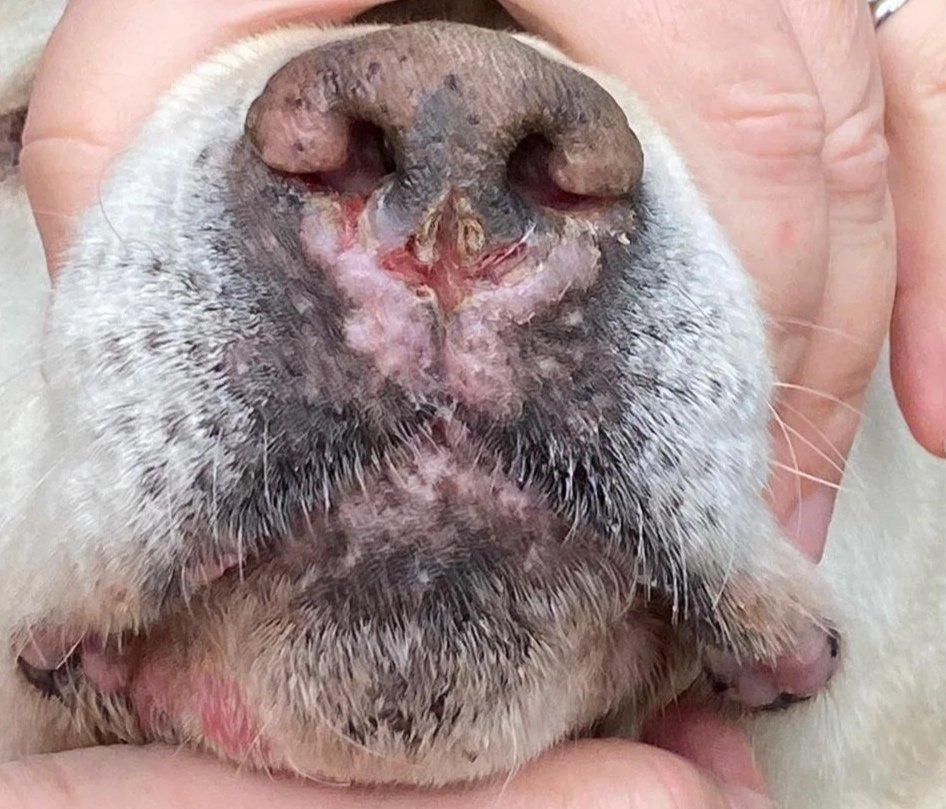
The dog with mucocutaneous disease
Sometimes the most important clue isn't on the trunk or limbs at all.
The disease may involve mucocutaneous junctions, including areas around the lips, nose, eyes, anus or genital region.
The owner may notice:
Redness
Loss of pigment
Scaling or crusting
Erosions
Ulceration
Changes around the lips or nose
Persistent lesions that don't heal normally
Mucocutaneous involvement is well recognised in canine epitheliotropic lymphoma and may occur alongside more widespread skin disease.
These lesions can have a surprisingly broad differential diagnosis, including infectious, autoimmune, inflammatory and neoplastic disease.
If treatment doesn't produce the expected response, histopathology becomes increasingly important.
What should owners look out for?
Owners don't need to recognise lymphoma themselves.
But there are some warning signs worth discussing with your veterinarian:
🚩 Skin disease that keeps coming back
Especially when treatment produces only temporary improvement.
🚩 Skin disease that keeps changing
For example, an area that starts as redness or scaling and gradually develops hair loss, thickening, plaques or ulceration.
🚩 Multiple unusual skin lesions
Particularly when they occur in several different locations.
🚩 Non-healing sores
A lesion that doesn't heal as expected deserves investigation.
🚩 Changes around the mouth, nose or other mucocutaneous areas
Persistent redness, scaling, pigment loss or ulceration should not automatically be assumed to be infection or allergy.
🚩 A diagnosis that doesn't quite fit
Sometimes the most important clue is simply that the patient isn't behaving like the diagnosis suggests it should.
What should veterinarians look out for?
Epitheliotropic lymphoma should enter the differential list when a patient has:
Chronic or recurrent dermatitis
Poor or incomplete response to appropriate therapy
Progressive alopecia and erythema
Multifocal plaques
Scaling or crusting without a convincing underlying cause
Hyperpigmentation or hypopigmentation
Persistent erosions or ulcers
Nodules or tumour-like lesions
Mucocutaneous involvement
Footpad lesions
Unusual or progressive skin disease in an older patient
How is epitheliotropic lymphoma diagnosed?
1. Detailed dermatological examination
Document:
Distribution
Primary and secondary lesions
Degree of alopecia
Erythema
Scaling
Crusting
Ulceration
Nodules or plaques
Mucocutaneous involvement
Footpads
Peripheral lymph nodes
2. Cytology
Cytology can be useful as an initial investigation and may identify an atypical lymphoid population.
However, a non-diagnostic cytology does not rule out epitheliotropic lymphoma.
The defining feature is where the neoplastic lymphocytes are located within the skin, something that generally requires histopathological examination.
3. Skin biopsy
This is often the critical diagnostic step.
Ideally, biopsy should target representative primary lesions, rather than heavily ulcerated or secondarily infected areas where possible.
Multiple biopsies from different types of lesions can be particularly helpful when the disease is heterogeneous.
Histopathology assesses the pattern of lymphocytic infiltration and, importantly, whether there is epitheliotropisminvolving the epidermis and/or adnexal structures.
4. Immunohistochemistry
Immunohistochemistry can help establish the phenotype of the neoplastic lymphocytes.
Most canine epitheliotropic cutaneous lymphomas are T-cell neoplasms.
CD3 is typically used to identify T cells, while markers such as CD20/CD79a can help evaluate B-cell populations.
This information can be important for confirming the diagnosis and understanding the biology of the disease.
Don't forget staging
Once lymphoma has been diagnosed, the next question is:
How extensive is the disease?
Depending on the individual patient, staging may include:
Complete physical examination
Peripheral lymph node assessment
CBC and serum biochemistry
Urinalysis
Thoracic imaging
Abdominal ultrasound
Assessment of liver and spleen
Lymph node sampling where indicated
Bone marrow evaluation in selected cases
Additional molecular testing where clinically relevant
A practical diagnostic checklist for veterinarian
History
☐ Duration and progression
☐ Previous diagnoses
☐ Response to treatment
☐ Degree of pruritus
☐ Previous corticosteroid use
☐ Antibiotic/antifungal response
☐ Development of new lesions
☐ Systemic signs
☐ Weight change
Examination
☐ Map lesion distribution
☐ Examine mucocutaneous junctions
☐ Examine footpads
☐ Palpate all peripheral lymph nodes
☐ Look for plaques/nodules
☐ Look for alopecia, erythema, scaling and ulceration
☐ Photograph representative lesions
Initial diagnostics
☐ Skin cytology
☐ Skin scrapings where indicated
☐ Appropriate infectious disease testing
☐ Bacterial/fungal culture where indicated
☐ CBC/biochemistry ± urinalysis
If disease is persistent, progressive or atypical
☐ Reconsider the differential diagnosis
☐ Obtain multiple skin biopsies
☐ Request dermatopathology review
☐ Specifically ask about epitheliotropism
☐ Consider immunohistochemistry
☐ Consider PARR/clonality testing
Once lymphoma is confirmed
☐ Determine immunophenotype
☐ Assess peripheral lymph nodes
☐ Stage appropriately
☐ Discuss prognosis
☐ Consider oncology referral
